Monday, March 27, 2017

Miscarriage & abortion

See also: Miscarriage and Abortion
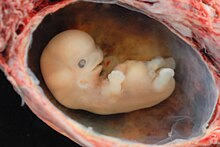
A complete spontaneous abortion at about 6 weeks from conception, i.e. 8 weeks from LMP
Some embryos do not survive to the next stage of development. When this happens naturally, it is called spontaneous abortion or miscarriage.[11] There are many reasons why this may occur. The most common natural cause of miscarriage is chromosomal abnormality in animals[12] or genetic load in plants.[13]
In species which produce multiple embryos at the same time, miscarriage or abortion of some embryos can provide the remaining embryos with a greater share of maternal resources. This can also disturb the pregnancy, causing harm to the second embryo. Genetic strains which miscarry their embryos are the source of commercial seedless fruits.
Abortion is the process of artificially (non-naturally) removing the embryo through deliberate pharmaceutical or surgical methods.

Research and technology

Embryos are used in various fields of research and in techniques of assisted reproductive technology. An egg may be fertilized in vitro and the resulting embryo may be frozen for later use. The potential of embryonic stem cell research, reproductive cloning, and germline engineering are currently being explored. Prenatal diagnosis or preimplantation diagnosis enables testing embryos for diseases or conditions.
Cryoconservation of animal genetic resources is a practice in which animal germplasms, such as embryos are collected and stored at low temperatures with the intent of conserving the genetic material.
The embryos of Arabidopsis thaliana have been used as a model to understand gene activation, patterning, and organogenesis of seed plants.[6]
In regards to research using human embryos, the ethics and legalities of this application continue to be debated.[7][8][9]

Fossilized embryos

Main article: Fossil embryos
Fossilized animal embryos are known from the Precambrian, and are found in great numbers during the Cambrian period. Even fossilized dinosaur embryos have been discovered.[10]

Development

In animals, the development of the zygote into an embryo proceeds through specific recognizable stages of blastula, gastrula, and organogenesis. The blastula stage typically features a fluid-filled cavity, the blastocoel, surrounded by a sphere or sheet of cells, also called blastomeres. In a placental mammal, an ovum is fertilized in a fallopian tube through which it travels into the uterus. An embryo is called a fetus at a more advanced stage of development and up until birth or hatching. In humans, this is from the eleventh week of gestation. However, animals which develop in eggs outside the mother's body, are usually referred to as embryos throughout development; e.g. one would refer to a chick embryo, not a "chick fetus," even at later stages.
During gastrulation the cells of the blastula undergo coordinated processes of cell division, invasion, and/or migration to form two (diploblastic) or three (triploblastic) tissue layers. In triploblastic organisms, the three germ layers are called endoderm, ectoderm, and mesoderm. The position and arrangement of the germ layers are highly species-specific, however, depending on the type of embryo produced. In vertebrates, a special population of embryonic cells called the neural crest has been proposed as a "fourth germ layer", and is thought to have been an important novelty in the evolution of head structures.
During organogenesis, molecular and cellular interactions between germ layers, combined with the cells' developmental potential, or competence to respond, prompt the further differentiation of organ-specific cell types.[citation needed] For example, in neurogenesis, a subpopulation of ectoderm cells is set aside to become the brain, spinal cord, and peripheral nerves. Modern developmental biology is extensively probing the molecular basis for every type of organogenesis, including angiogenesis (formation of new blood vessels from pre-existing ones), chondrogenesis (cartilage), myogenesis (muscle), osteogenesis (bone), and many others.

Plant embryos

Further information: Sporophyte
The inside of a Ginkgo seed, showing the embryo
In botany, a seed plant embryo is part of a seed, consisting of precursor tissues for the leaves, stem (see hypocotyl), and root (see radicle), as well as one or more cotyledons. Once the embryo begins to germinate — grow out from the seed — it is called a seedling (plantlet).
Bryophytes and ferns also produce an embryo, but do not produce seeds. In these plants, the embryo begins its existence attached to the inside of the archegonium on a parental gametophyte from which the egg cell was generated. The inner wall of the archegonium lies in close contact with the "foot" of the developing embryo; this "foot" consists of a bulbous mass of cells at the base of the embryo which may receive nutrition from its parent gametophyte. The structure and development of the rest of the embryo varies by group of plants. Once the embryo has expanded beyond the enclosing archegonium, it is no longer termed an embryo.

Delivery

For a Morpholino to be effective, it must be delivered past the cell membrane into the cytosol of a cell. Once in the cytosol, Morpholinos freely diffuse between the cytosol and nucleus, as demonstrated by the nuclear splice-modifying activity of Morpholinos observed after microinjection into the cytosol of cells. Different methods are used for delivery into embryos, into cultured cells or into adult animals. A microinjection apparatus is usually used for delivery into an embryo, with injections most commonly performed at the single-cell or few-cell stage;[37] an alternative method for embryonic delivery is electroporation, which can deliver oligos into tissues of later embryonic stages.[38] Common techniques for delivery into cultured cells include the Endo-Porter peptide (which causes the Morpholino to be released from endosomes),[14] the Special Delivery system (no longer commercially available, used a Morpholino-DNA heteroduplex and an ethoxylated polyethylenimine delivery reagent),[13] electroporation,[39] or scrape loading.[40]
Delivery into adult tissues is usually difficult, though there are a few systems allowing useful uptake of unmodified Morpholino oligos (including uptake into muscle cells with Duchenne muscular dystrophy[41] or the vascular endothelial cells stressed during balloon angioplasty[42]). Though they permeate through intercellular spaces in tissues effectively, unconjugated PMOs have limited distribution into the cytosol and nuclear spaces within healthy tissues following IV administration. Systemic delivery into many cells in adult organisms can be accomplished by using covalent conjugates of Morpholino oligos with cell-penetrating peptides, and, while toxicity has been associated with moderate doses of the peptide conjugates,[43][44] they have been used in vivo for effective oligo delivery at doses below those causing observed toxicity.[5][45] An octa-guanidinium dendrimer attached to the end of a Morpholino can deliver the modified oligo (called a Vivo-Morpholino) from the blood to the cytosol.[15][46] Delivery-enabled Morpholinos, such as peptide conjugates and Vivo-Morpholinos, show promise as therapeutics for viral and genetic diseases.[47]

Specificity, stability and non-antisense effects

Morpholinos have become a standard knockdown tool in animal embryonic systems, which have a broader range of gene expression than adult cells and can be strongly affected by an off-target interaction. Following initial injections into frog or fish embryos at the single-cell or few-cell stages, Morpholino effects can be measured up to five days later,[31] after most of the processes of organogenesis and differentiation are past, with observed phenotypes consistent with target-gene knockdown. Control oligos with irrelevant sequences usually produce no change in embryonic phenotype, evidence of the Morpholino oligo's sequence-specificity and lack of non-antisense effects. The dose required for a knockdown can be reduced by coinjection of several Morpholino oligos targeting the same mRNA, which is an effective strategy for reducing or eliminating dose-dependent off-target RNA interactions.[32]
MRNA rescue experiments can often restore the wild-type phenotype to the embryos and provide evidence for the specificity of a Morpholino. In an mRNA rescue, a Morpholino is co-injected with an mRNA that codes for the morphlino's protein. However, the rescue mRNA has a modified 5'-UTR (untranslated region) so that the rescue mRNA contains no target for the Morpholino. The rescue mRNA's coding region encodes the protein of interest. Translation of the rescue mRNA replaces production of the protein that was knocked down by the Morpholino. Since the rescue mRNA would not affect phenotypic changes due to the Morpholino's off-target gene expression modulation, this return to wild-type phenotype is further evidence of Morpholino specificity.[31]
Because of their completely unnatural backbones, Morpholinos are not recognized by cellular proteins. Nucleases do not degrade Morpholinos,[33] nor are they degraded in serum or in cells.[34] Morpholinos do not activate toll-like receptors or innate immune responses such as interferon induction or the NF-κB-mediated inflammation response. Morpholinos are not known to modify DNA methylation.
Up to 18% of Morpholinos appear to induce nontarget-related phenotypes including cell death in the central nervous system and somite tissues of zebrafish embryos.[35] Most of these effects are due to activation of p53-mediated apoptosis and can be suppressed by co-injection of an anti-p53 Morpholino along with the experimental Morpholino. Moreover, the p53-mediated apoptotic effect of a Morpholino knockdown has been phenocopied using another antisense structural type, showing the p53-mediated apoptosis to be a consequence of the loss of the targeted protein and not a consequence of the knockdown oligo type.[36] It appears that these effects are sequence-specific; as in most cases, if a Morpholino is associated with non-target effects, the 4-base mismatch Morpholino will not trigger these effects.
A cause for concern in the use of Morpholinos is the potential for "off-target" effects. Whether an observed morphant phenotype is due to the intended knockdown or an interaction with an off-target RNA can often be addressed by running another experiment to confirm that the observed morphant phenotype results from the knockdown of the expected target. This can be done by recapitulating the morphant phenotype with a second, non-overlapping Morpholino targeting the same mRNA[31] or by confirmation of the observed phenotypes by use of a mutant strain or dominant-negative methods. As mentioned above, rescue of observed phenotypes by coinjecting a rescue mRNA is, when feasible, a reliable test of specificity of a Morpholino.[31]

Other applications: blocking other mRNA sites and use as probes

Other applications: blocking other mRNA sites and use as probes

Morpholinos have been used to block miRNA activity[23][24] and maturation.[25] Fluorescein-tagged Morpholinos combined with fluorescein-specific antibodies can be used as probes for in-situ hybridization to miRNAs.[26] Morpholinos can block ribozyme activity.[27] U2 and U12 snRNP functions have been inhibited by Morpholinos.[28] Morpholinos targeted to "slippery" mRNA sequences within protein coding regions can induce translational frameshifts.[29] Morpholinos can block RNA editing.[30] Morpholino activities against this variety of targets suggest that Morpholinos can be used as a general-purpose tool for blocking interactions of proteins or nucleic acids with mRNA.

Normal gene expression in eukaryotes

See also: Gene expression
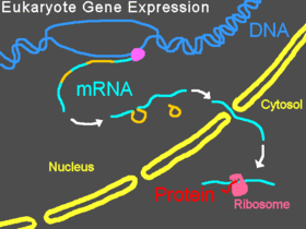
Eukaryotic gene expression without intervention by a Morpholino
In eukaryotic organisms, pre-mRNA is transcribed in the nucleus, introns are spliced out, then the mature mRNA is exported from the nucleus to the cytoplasm. The small subunit of the ribosome usually starts by binding to one end of the mRNA and is joined there by various other eukaryotic initiation factors, forming the initiation complex. The initiation complex scans along the mRNA strand until it reaches a start codon, and then the large subunit of the ribosome attaches to the small subunit and translation of a protein begins. This entire process is referred to as gene expression; it is the process by which the information in a gene, encoded as a sequence of bases in DNA, is converted into the structure of a protein. A Morpholino can modify splicing or block translation, depending on the Morpholino's base sequence.

Blocking translation

Translation blocked by a Morpholino oligo
Bound to the 5'-untranslated region of messenger RNA (mRNA), Morpholinos can interfere with progression of the ribosomal initiation complex from the 5' cap to the start codon. This prevents translation of the coding region of the targeted transcript (called "knocking down" gene expression). This is useful experimentally when an investigator wishes to know the function of a particular protein; Morpholinos provide a convenient means of knocking down expression of the protein and learning how that knockdown changes the cells or organism. Some Morpholinos knock down expression so effectively that, after degradation of preexisting proteins, the targeted proteins become undetectable by Western blot.[16]
In 2016 a synthetic peptide-conjugated PMO (PPMO) was found to inhibit the expression of New Delhi Metallo-beta-lactamase, an enzyme that many drug-resistant bacteria use to destroy carbapenems.[17][18]

Modifying pre-mRNA splicing

Splicing blocked by a Morpholino oligo
Morpholinos can interfere with pre-mRNA processing steps either by preventing splice-directing small nuclear ribonucleoproteins (snRNP) complexes from binding to their targets at the borders of introns on a strand of pre-mRNA, or by blocking the nucleophilic adenine base and preventing it from forming the splice lariat structure, or by interfering with the binding of splice regulatory proteins such as splice silencers[19] and splice enhancers.[20] Preventing the binding of snRNP U1 (at the donor site) or U2/U5 (at the polypyrimidine moiety and acceptor site) can cause modified splicing, commonly excluding exons from the mature mRNA. Targeting some splice targets results in intron inclusions, while activation of cryptic splice sites can lead to partial inclusions or exclusions.[21] Targets of U11/U12 snRNPs can also be blocked.[22] Splice modification can be conveniently assayed by reverse-transcriptase polymerase chain reaction (RT-PCR) and is seen as a band shift after gel electrophoresis of RT-PCR products.[3]